Flow cytometry is a technology that allows the analysis of cells, particles, or microorganisms as they pass through one or more lasers while suspended in a buffered saline solution. Each particle is analysed for visible light scattering and one or multiple fluorescence parameters; therefore, it must be possible to label the particles with one or more fluorescent substances (eg, fluorescence-conjugated antibodies, propidium iodide, or green fluorescent protein (GFP), etc). This multiparametric study allows analysing of subpopulations in complex samples.
The recently created IISPV Flow Cytometry Facility is dedicated to helping both internal and external IISPV scientists with technical-scientific advice to develop reliable flow cytometry assays that meet current quality standards.
With the collaboration of:
Acquisition and Analysis: Study of cells or particles based on their fluorescent or light scattering characteristics. Among the acquisition services offered are:
Cell Sorting: FACS Aria III is an analyzer and separator cytometer that allows cells or particles to be separated based on parameters previously defined by the operator.
Advice and experimental design: The Facility offers advice to users on experimental design, upon request of the principal investigator and according to the availability of the Unit’s staff.
Self-service: The self-service regime is targeted towards advanced users, previously trained and authorized by UCF.
Training: The Flow Cytometry Facility has started a series of training courses aimed at higher education and university students, specifically within the URV.
Analysis of Results: The Facility makes available to users a license of the FlowJo flow cytometry analysis program, which allows offline analysis of the results. In addition, the FACS Aria III and Cytek Northern Lights equipment includes the BD FACS DIVA™ and SpectroFlo analysis software, respectively.
Flow cytometers analyzers
Cell sorters (flow cytometer)
Nanoparticle characterization systems
Other cell separation systems
Additional laboratory resources
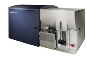
FACS Aria III Flow Cytometer and Cell Sorter (BD Biosciences)
The FACS Aria III (BD) is a high-speed flow cytometer that combines multiparametric analysis with cell sorting capabilities. It is equipped with 3 lasers:
(See: Equipment configuration).
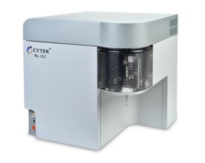
Northern Lights Spectral Flow Cytometer (Cytek®)
The Northern Lights (Cytek®) is a flow cytometer based on spectral technology, enabling greater resolution and flexibility in multicolour panel design.
It is equipped with three lasers:
The system includes 41 detectors and can capture up to 38 spectral channels. Unlike conventional flow cytometry, this platform records the full emission spectrum of each fluorochrome, generating a unique spectral signature for every marker.
This technology significantly enhances the identification of complex cell populations, especially in samples with high autofluorescence or when using high-dimensional panels.
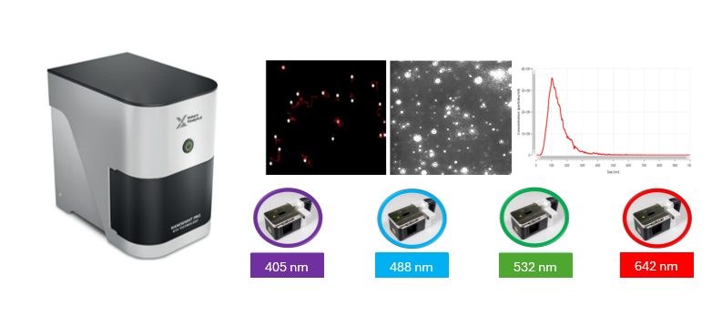
NSPro Nanoparticle Analyzer (IESMAT – NTA)
The NSPro (IESMAT) is an advanced analysis system based on Nanoparticle Tracking Analysis (NTA) technology, designed for the precise characterization of particles in suspension ranging from 10 nm to 2 μm. This size range includes extracellular vesicles, exosomes, viruses, and synthetic nanoparticles.
The instrument uses a laser to illuminate the sample and a high-sensitivity camera that captures the Brownian motion of particles in real time. Based on the individual particle displacement, size and concentration are calculated using the Stokes–Einstein equation.
The system includes a fluorescence module equipped with long-pass filters and excitation lasers at 405 nm, 488 nm, 532 nm, and 642 nm, allowing the use of one fluorochrome or fluorescent probe per experiment. This configuration is well-suited for studies involving labeled particles such as extracellular vesicles or functionalized nanoparticles.
This technology provides detailed information on particle size distribution and concentration, enabling the characterization of small particles with applications in nanomedicine, cell biology, and the development of advanced therapies.
Erika Scholz, PhD. Head of Flow Cytometry and Cell Sorting Facility.
A/e: erika.scholz@irbcatsud.cat
Orcid: https://orcid.org/0000-0002-7589-0517
Oficina técnica IISPV / Planta 7 del Hospital Universitario Joan XXIII
Institut d’Investigació Sanitària Pere Virgili (IISPV)
Hospital Universitari Joan XXIII C/ Doctor Mallafrè Guasch, 4
43005 Tarragona
Telèfon: +34 977 297 996
The Cell Culture Platform is a technical and scientific support unit that offers support to research groups, both internal and external to the IISPV, providing them with the appropriate facilities and equipment for the cultivation, maintenance and execution of bioassays with eukaryotic cells.


The Statistical Support Platform is a cross-cutting platform for statistical support to researchers that offers a support and advice service in all phases of the development of a research project: methodology, design, analysis and publication.
The Clinical Pharmacy Department is a central clinical support service with its own activities and support for the other healthcare services of the ICS Camp de Tarragona.
Bioinformatics service available to all IISPV professionals.
The Cell Culture Platform is a technical and scientific support unit that offers support to research groups, both internal and external to the IISPV, providing them with the appropriate facilities and equipment for the cultivation, maintenance and execution of bioassays with eukaryotic cells.
The Statistical Support Platform is a cross-cutting platform for statistical support to researchers that offers a support and advice service in all phases of the development of a research project: methodology, design, analysis and publication.
The Clinical Pharmacy Department is a central clinical support service with its own activities and support for the other healthcare services of the ICS Camp de Tarragona.
Bioinformatics service available to all IISPV professionals.


