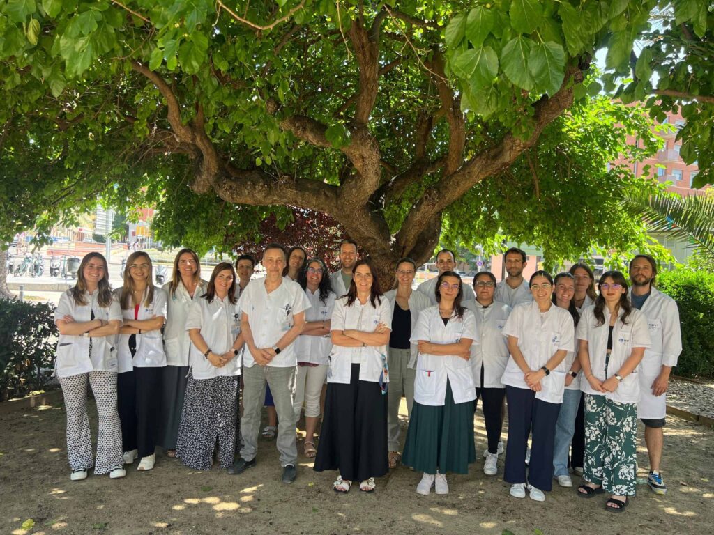
The head of the Hepatology Research Group at IRB CatSud, Dr. Jordi Gracia-Sancho, took part as an invited speaker at the International Congress on Coagulopathy in Liver Disease, held in Castellana Grotte (Italy) from 8 to 10 April. The international meeting focused on the mechanisms of coagulopathy associated with liver disease.
In this context, Dr. Gracia-Sancho delivered the opening lecture of the congress, centred on the development of endothelial dysfunction in chronic liver disease. His presentation placed special emphasis on the molecular and cellular mechanisms contributing to endothelial alteration —which regulates blood flow— as well as on the key role of the hepatic vascular compartment in disease progression.
Another topic addressed at the International Congress on Coagulopathy in Liver Disease was the emerging therapeutic opportunities aimed at restoring endothelial function and modulating hepatic microcirculation, with the goal of slowing disease progression and promoting its regression. The lecture highlighted the importance of the endothelium as a therapeutic target in liver pathology.
A new study reveals relevant muscular and metabolic alterations in patients with long COVID
A research team from the Research Group on Autoimmunity, Infection and Thrombosis (GRAIÏT) at the Southern Catalonia Biomedical Research Institute (formerly IISPV), from at the same time of the Internal Medicine and Clinical Physiology and Functional Evaluation services at the Sant Joan University Hospital in Reus, part of the has presented new evidence confirming the deep and lasting impact of SARS‑CoV‑2 infection on the muscular and metabolic systems of individuals affected by Post‑COVID‑19 Condition (long COVID). The results of the study “Assessment of Physical Status and Analysis of Lipidomic and Metabolomic Alterations in Patients with Post‑COVID‑19 Condition” highlight the need to promote specific rehabilitation and physical reconditioning programs to improve patient recovery, and have been published in the scientific journal PLOS ONE this March.
The study reveals significant peripheral muscle involvement related to the infection. This alteration leads to a marked loss of physical performance, one of the most frequent and disabling symptoms of the condition. The researchers emphasize that patients may benefit from personalized rehabilitation protocols to regain strength, endurance, and functionality.
Additionally, through the analysis of body metabolism using metabolomic and lipidomic techniques, they detected changes that may explain some of the patients’ symptoms:
According to the study’s authors, these findings reinforce the idea that Post‑COVID‑19 Condition is a multisystemic disorder requiring an integrated approach combining physical rehabilitation, metabolic monitoring, and personalized interventions.
The researchers note that the results open the door to developing more specific therapeutic strategies and to better guiding care pathways for patients with long COVID.
Schizophrenia is a complex neuropsychiatric disorder in which genetics plays a key role. A recent study focuses on the so‑called copy number variants, which are DNA fragments that, instead of having the usual amount, show duplications or — most relevant in this case — losses of genetic material.
A team of researchers from the Hospital Universitari Institut Pere Mata (HUIPM), the Institut de Recerca Biomèdica Catalunya Sud (IRB CatSud), and Rovira i Virgili University (URV) has led a study that sheds new light on why schizophrenia may appear earlier in some individuals than in others. The research, published in the scientific journal Schizophrenia Research, indicates that the loss of genetic material may influence the age at which the disorder manifests.
The study analyzes 836 participants — 323 with schizophrenia and 513 without — and focuses on a type of DNA alteration that includes small losses of genetic material. The findings emphasize that it is specifically these losses — and not duplications — that are linked to an earlier onset of the disease. Such losses can advance the onset of schizophrenia and are not harmless, as they may eliminate entire genes or regulatory elements that control how these genes function, potentially disrupting the balance required for proper brain development.
Researchers observed that people with schizophrenia show a higher overall burden of variations in DNA fragment quantity compared with healthy participants. This result reinforces the idea that structural alterations across our entire DNA play an important role in the development of the disorder.
Losses of genetic material are associated with an earlier onset. One of the key findings of the study is that the number of missing DNA fragments is related to the age at which schizophrenia begins. “Specifically, individuals with a greater lack of DNA fragments tend to develop symptoms earlier, while patients with later onset present levels similar to those of people without the disorder. This suggests that, beyond whether someone will develop schizophrenia, the total amount of copy number alterations could influence the age of onset,” explains the study’s lead researcher, Gerard Muntané.
Although each individual variation has a modest effect, the cumulative impact may influence the onset of the disease at the population level. The authors emphasize the need for larger and more diverse samples to confirm these results and to better understand the biological mechanisms involved in schizophrenia. This would allow earlier identification of patients at higher risk of early onset and guide strategies for early detection and personalized intervention.
Article reference
Muntané, G., Valle, A., Ramon-Cañellas, P., Martorell, L., & Vilella, E. (2026). The impact of CNV burden on age at onset of schizophrenia. Schizophrenia Research, 291, 12–19. https://doi.org/10.1016/j.schres.2026.02.006
A study by IRB Catalunya Sud (formerly IISPV) and Hospital Universitari Joan XXIII de Tarragona has discovered that these vesicles act differently depending on tumour aggressiveness, opening new possibilities for future therapeutic strategies.
Prostate cancer is the most common tumour in men in many Western countries. In the Tarragona region, nearly 670 new cases are diagnosed each year, and the number exceeds 30,000 at the national level. Although many tumours grow slowly, others can progress and spread, so understanding the factors that drive this aggressiveness is essential to improve patient outcomes.
In this context, researchers from the Grup de Recerca en Biomarcadors de Malalties i Mecanismes Moleculars (DIBIOMEC) at the Institut de Recerca Biomèdica Catalunya Sud (IRB Catalunya Sud, formerly IISPV), in collaboration with the Urology and Pathology Departments of Hospital Universitari Joan XXIII de Tarragona, have made an important step forward. Their recently published study shows for the first time that extracellular vesicles (small particles released by cells) from the adipose tissue surrounding the prostate (periprostatic adipose tissue, PPAT) modulate the behaviour of tumour cells differently depending on cancer risk level.
The research, led by Dra. Matilde R. Chacón and Dr. Xavier Ruiz-Plazas, and carried out by a multidisciplinary team, provides a new perspective on how the tumour microenvironment — particularly periprostatic fat — “communicates” with cancer and influences its evolution.
First, the study reveals risk‑dependent effects. Vesicles from periprostatic adipose tissue of patients with low‑risk prostate cancer mainly stimulate tumour cell proliferation. In contrast, vesicles from patients with high‑risk tumours do not promote proliferation, but they do increase the migration capacity of cancer cells and stimulate angiogenesis (the formation of new blood vessels), both of which are key processes in tumour progression and spread.
The vesicles also affect the tumour microenvironment. Besides acting on cancer cells, they influence other surrounding cells. Low‑risk vesicles promote a pro‑inflammatory and immunosuppressive profile in macrophages (immune cells), which may help create a supportive environment for early‑stage tumours.
Another important finding is the activation of signalling pathways, as the observed effects are linked to the activation of key molecular routes in cancer.
This discovery shows that periprostatic adipose tissue is not just a passive structure but an active and dynamic player that modulates prostate cancer behaviour depending on disease aggressiveness. Extracellular vesicles from this tissue emerge as promising therapeutic targets for future strategies aimed at interfering with communication between the tumour and its microenvironment, especially in cases with a higher risk of progression.
Although these results come from in vitro models and further studies will be needed in more complex systems, this research represents a significant step forward in understanding prostate cancer biology and opens a promising avenue for translational research.
Link to the scientific publication:
Arreaza-Gil V. et al. Periprostatic adipose tissue-derived extracellular vesicles modulate prostate cancer cell behaviour in vitro according to tumour grade. Mol Med (2026).
PubMed: https://pubmed.ncbi.nlm.nih.gov/41566212/
DOI: 10.1186/s10020-026-01422-7
The Universitat Rovira i Virgili (URV), with the participation of the Institut de Recerca Biomèdica Catalunya Sud (IRB CatSud, formerly IISPV), has taken part in a pioneering study showing that consuming extra virgin olive oil may help preserve cognitive function by modulating the gut microbiota. The research, published in Microbiome, is the first study in humans to analyse this specific relationship.
The study was carried out using data from 656 people aged 55 to 75 with overweight or obesity and metabolic syndrome, all participating in the PREDIMED-Plus project. Participants who consumed extra virgin olive oil —and not refined oil— showed a better evolution of cognitive function and a more diverse gut microbiota, which is a key indicator of metabolic health. In addition, the bacterial genus Adlercreutzia was identified as a possible mediator of this protective effect.
The difference between the two types of olive oil lies in the production process: while extra virgin olive oil preserves antioxidants, polyphenols and bioactive compounds, refined oil loses most of these elements during industrial processing. “Not all olive oils have the same benefits for cognitive function,” explains Jiaqi Ni, first author of the study.
The results highlight the importance of fat quality within the Mediterranean diet. “Extra virgin olive oil not only protects the heart, but may also help preserve the brain during ageing,” says Jordi Salas-Salvadó, principal investigator. Codirectors Nancy Babio and Stephanie Nishi emphasise that, in a context of increasing cognitive decline, improving diet quality is an accessible and effective strategy.
This research was made possible thanks to the leadership of the URV and IISPV-CERCA, with the collaboration of CIBERobn and international institutions such as Wageningen University and Harvard University.
A study led by the Diabetes and Associated Metabolic Diseases (DIAMET) group at the Institut de Recerca Biomèdica Catalunya Sud (IRB CatSud, formerly IISPV) shows that liver fibrosis and type 2 diabetes significantly modify the hormonal response after eating in people with fatty liver associated with metabolic dysfunction (MASLD). The research, published in the Journal of Physiology and Biochemistry, examines how both conditions affect the secretion of essential hormones for glucose control—such as glucagon and the incretins (GLP‑1, GLP‑2 and GIP)—after a standardized meal.
The results show that liver fibrosis is the most important factor influencing the increase in GLP‑1 levels, both in fasting conditions and after the meal, regardless of whether the patient has diabetes. In addition, when liver fibrosis and type 2 diabetes occur together, hormonal changes become stronger, suggesting a synergistic effect between both conditions. Type 2 diabetes is also linked to the loss of the normal suppression of glucagon after eating, a key process to keep blood glucose within healthy ranges.
These findings reinforce the idea that a fibrotic liver is not a passive organ, but plays an active role in metabolic dysregulation. Fibrosis not only reflects past damage but also contributes to generating new hormonal alterations. Understanding these changes is essential to improve clinical assessment and move towards more personalised treatments for MASLD.
With the growing prevalence of MASLD and type 2 diabetes, understanding how these conditions interact at the hormonal level is crucial to improve early diagnosis and optimise treatments based on the incretin–glucagon axis. The study provides evidence that can help identify subgroups of patients who may benefit from more specific therapeutic approaches, with a direct impact on clinical practice.
Overall, the results confirm that liver fibrosis is a central determinant of GLP‑1 levels, and that the coexistence of type 2 diabetes further intensifies these hormonal alterations. This knowledge highlights the need to design therapeutic strategies adapted to the metabolic and liver profile of each patient, especially in a context where both diseases are becoming increasingly common.
The study involved researchers from the Universitat Rovira i Virgili (URV), the Institut d’Investigacions Biomèdiques August Pi i Sunyer (IDIBAPS) – Hospital Clínic Barcelona, the research networks of the Instituto de Salud Carlos III (ISCIII) — the Centro de Investigación Biomédica en Red de Diabetes y Enfermedades Metabólicas Asociadas (CIBERDEM) and the Centro de Investigación Biomédica en Red de Enfermedades Hepáticas y Digestivas (CIBEREHD) — as well as the Hospital Universitari Joan XXIII (Tarragona).
The grants, with more than €270,000 in funding, support advanced projects in personalized radiotherapy and artificial intelligence. A third grant is aimed at specialized training in molecular oncology
The Spanish Association Against Cancer in Tarragona presented on Thursday, 19 February, the three grants awarded for the 2025 call. These grants show a strong commitment to cancer research and to the goal of reaching a 70% cancer survival rate by 2030.
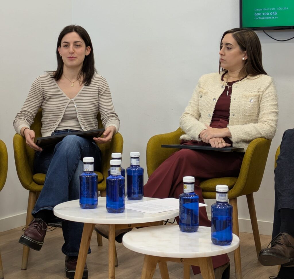
Two of the funded researchers work at the Institute of Biomedical Research of Southern Catalonia (IRB CatSud, formerly IISPV). They lead projects that combine technology, innovation, and clinical impact: Dr Bárbara Antonia Malavé and Marta Canela.
Dr Bárbara Antonia Malavé has received the Clinic Junior Grant AECC 2025, supporting a project focused on improving radiotherapy treatment for prostate cancer. The project uses advanced biomarkers and artificial intelligence to adapt treatments to each patient. The grant provides €154,000 over four years.
Also at IRB CatSud, Marta Canela has received the AECC Tarragona Predoctoral Grant 2025. Her research combines medical imaging and blood analysis to predict how patients with lung cancer will respond to radiotherapy. The grant provides €110,660 over four years.
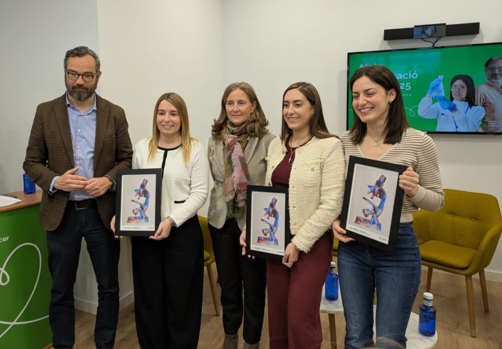
The third grant has been awarded to Maria Guirro, who will receive the Clinic Training Grant AECC 2025. The €7,100 contribution will allow her to complete the Master’s Degree in Molecular Oncology (MOM).
A study of 746 older people followed for six years identifies a “microbial signature” associated with this dietary pattern and more favourable cognitive ageing
Following a Mediterranean diet not only benefits the heart and metabolism, but could also help preserve cognitive function as ageing progresses. This is according to research led by the Rovira i Virgili University (URV), the IISPV and CIBERobn, which shows how this dietary pattern is associated with a healthier gut microbiota and slower cognitive decline in older adults with overweight or obesity and metabolic syndrome.
The study, published in the journal BMC Medicine, analysed data from 746 older adults at high cardiometabolic risk, who were followed for six years. The research team assessed adherence to the Mediterranean diet, the composition of the gut microbiota and the progression of cognitive function over time. The results indicate that those who most faithfully followed this dietary pattern had a more favourable gut microbiota and a more positive cognitive trajectory.
One of the most innovative aspects of the work is the identification of a “microbial fingerprint” characteristic of the Mediterranean diet. This new biomarker, based on the presence and abundance of certain gut bacteria associated with this type of diet, is also linked to slower cognitive decline. According to the authors, this finding provides new clues about the biological mechanisms that explain the benefits of the Mediterranean diet on the brain.
Eix intestí-cervell
Cognitive function includes abilities such as memory, attention, learning, language and decision-making, which are essential for maintaining autonomy in everyday life. In parallel, the gut microbiota is composed of trillions of bacteria that are involved in key processes such as digestion, immunity and the production of substances that influence the body’s functioning. In recent years, research has revealed the existence of the so-called gut-brain axis, a bidirectional communication system through which intestinal microorganisms can produce compounds that reach the brain and affect its functioning.
“Aquest estudi demostra que la microbiota intestinal és una peça clau en els beneficis cognitius de la dieta mediterrània”, explica Jiaqi Ni, primera autora del treball i investigadora predoctoral de la URV. “Els nostres resultats suggereixen que alguns bacteris intestinals associats a una major adherència a aquest patró alimentari podrien protegir davant del deteriorament cognitiu”.
En la mateixa línia, el catedràtic de la URV Jordi Salas-Salvadó, director de l’estudi, destaca que “identificar una empremta microbiana associada a la dieta mediterrània obre noves oportunitats per dissenyar intervencions nutricionals o microbianes orientades a promoure un envelliment cognitiu saludable”. De la seva banda, les investigadores del Departament de Bioquímica i Biotecnologia de la URV Nancy Babio i Stephanie K. Nishi subratllen la rellevància dels resultats en un context d’envelliment poblacional i augment de la prevalença de la demència, i apunten que millorar la qualitat de la dieta és una estratègia senzilla i accessible amb beneficis reals per a la salut cerebral.
El treball ha estat liderat per la investigadora predoctoral Jiaqi Ni i dirigit per Jordi Salas-Salvadó, Nancy Babio i Stephanie K. Nishi, membres de la Unitat de Nutrició Humana del Departament de Bioquímica i Biotecnologia de la URV, amb la col·laboració d’investigadors del consorci PREDIMED-Plus. L’estudi s’emmarca en una recerca multicèntrica que contribueix a aprofundir en la relació entre alimentació, microbiota intestinal i salut cerebral al llarg de l’envelliment.
Referència bibliogràfica: Ni J, Hernández-Cacho A, Nishi SK, Babio N, Belzer C, Konstati P, Vioque J, Corella D, Castañer O, Vidal J, Moreno-Indias I, Torres-Collado L, Coltell O, Fitó M, Ruiz-Canela M, Wang DD, Tinahones FJ, Salas-Salvadó J. Mediterranean diet, gut microbiota, and cognitive decline in older adults with obesity/overweight and metabolic syndrome: a prospective cohort study. BMC Med. 2025 Dec 1;23(1):669. doi: 10.1186/s12916-025-04488-y.
Maternal nutrition is essential for babies’ cognitive development, but some nutrients have received less attention in scientific research. Vitamin K, mainly known for its role in blood clotting, is one example. Although it is found in leafy green vegetables and vegetable oils, and is involved in processes related to glucose metabolism, insulin, and antioxidant and anti-inflammatory activity, its possible influence on children’s neurological development had not been explored until now.

A new study led by teams from the Universitat Rovira i Virgili (URV) and IRB CatSud (formerly IISPV), in collaboration with ISGlobal, provides the first evidence linking maternal intake of vitamin K1 (phylloquinone) with children’s neurodevelopment.
The research was directed by Mònica Bulló, head of the Nutrition and Metabolic Health group at URV and the TecnATox research center, and by Jordi Júlvez, researcher at IRB CatSud. The results have been published in the scientific journal Pediatric Research.
The study analysed 1,080 pregnant women and their children from the BiSC cohort. To estimate vitamin K intake, participants completed a food frequency questionnaire with 114 items. Later, several areas of early childhood neurodevelopment —cognition, socioemotional and physical development, communication, and behaviour— were evaluated at different stages.
The data show that higher vitamin K intake during pregnancy is associated with better overall development in children, as well as specific improvements in cognitive abilities and physical development. The study also suggests that doubling the current vitamin K recommendations for the general population could be linked to better communication skills and expressive language. Although this is an observational study and cannot prove direct causality, the research team believes it opens new paths to deepen our understanding of the role of maternal nutrition in neurocognitive development.
“The evidence we found suggests that vitamin K may play a beneficial role, and this could lead to future dietary recommendations specifically for pregnant women, with the aim of promoting optimal cognitive development in early childhood,” explains URV professor Mònica Bulló.
Bibliographic reference:
Mateu-Fabregat, J., Panisello, L., Novau-Ferré, N. et al. Maternal dietary phylloquinone intake (vitamin K1) and early childhood neurodevelopment. Pediatr Res (2025). https://doi.org/10.1038/s41390-025-04543-7
A study from URV, with participation from IISPV, compares abdominal ultrasound with magnetic resonance imaging and shows the potential of this technique to detect early risk of prediabetes and metabolic syndrome in people with abdominal obesity
The distribution of body fat, especially the fat accumulated in the abdomen, is a key factor in the risk of developing metabolic and cardiovascular diseases. However, not all abdominal fat has the same impact on health: subcutaneous fat, located under the skin, does not carry the same risks as visceral fat, which is stored deeper and can come into direct contact with vital organs. Identifying which type of fat is more predominant in a person is essential to assess metabolic risk and to guide clinical interventions. In this context, a study led by researchers from the Department of Medicine and Surgery at the Universitat Rovira i Virgili (URV), with the participation of research staff from the IISPV, analysed how useful and reliable abdominal ultrasound can be for measuring visceral fat, compared with magnetic resonance imaging, which is currently considered the reference technique.
In clinical practice, the risk associated with abdominal fat is usually assessed by measuring waist circumference. However, this simple method is limited because it cannot distinguish between subcutaneous and visceral fat. Imaging techniques such as magnetic resonance imaging or computed tomography provide this information with high accuracy, but they are expensive, require specialised equipment, and are not easily available in primary care. To explore more accessible alternatives, the research team aimed to validate the use of abdominal ultrasound to characterise fat distribution. “Ultrasound is available in most primary care centres and hospitals, and with trained health professionals it allows real-time imaging at a very low cost,” explains Claudia Jiménez-ten Hoevel, researcher at the Department of Medicine and Surgery at the URV and co-author of the article.
The key question was how similar the results of ultrasound could be to those of magnetic resonance imaging when analysing abdominal fat. To answer this, the team worked with a sample of 113 adult volunteers with abdominal obesity, living in Reus and nearby areas. All participants underwent both a magnetic resonance imaging scan and an abdominal ultrasound within a short period —between three and four days— to ensure comparable results.
A valid and accurate tool
The results of the study, published in Diabetes, Obesity and Metabolism, show that visceral fat measurements obtained through ultrasound had good agreement with those obtained through magnetic resonance imaging. However, the researchers also found some limitations of ultrasound when measuring subcutaneous fat, something that previous studies had already suggested. “The main conclusion is that ultrasound can be especially useful when the goal is to identify visceral fat, which is the type most clearly linked to metabolic risk,” says Anna Pedret, researcher from the same department. This potential adds to the fact that ultrasound is easy to use for trained staff and is available in most health centres.
Visceral fat: an indicator of metabolic risk
The analysis also allowed the researchers to explore whether the amount of visceral fat measured by ultrasound could predict future metabolic problems. The results show a relationship between high levels of visceral fat and the presence of prediabetes —a condition where blood sugar levels are high but still below the threshold for type 2 diabetes— as well as metabolic syndrome —a group of risk factors that increase the likelihood of cardiovascular disease.
More specifically, the study identifies a level of visceral fat above which the risk of associated diseases increases significantly. For example, a thickness of 7.35 centimetres or more is linked to a higher risk of prediabetes, while from 5.77 centimetres there is a relationship with the probability of metabolic syndrome. These findings highlight the potential of ultrasound as an exploratory tool: “Including abdominal ultrasound in routine clinical practice, especially in nutrition and primary care, could improve risk assessment and allow more personalised interventions,” the researchers explain.
The research group Nutrició Funcional, Oxidació i Malalties Cardiovasculars (NFOC-SALUT) from the URV and the Institut d’Investigació Sanitària Pere Virgili (IISPV) continues working to promote the use of ultrasound as an affordable, fast, and easily applicable technique in clinical practice. The team is now studying the impact of abdominal fat distribution in other population groups and its relationship with different health conditions, such as cognitive function or sarcopenia. In this context, the group is carrying out new studies using abdominal ultrasound in people aged 60 to 74, with ongoing research and open participation. Interested individuals can contact the team at estudisalimentacionfocsalut@gmail.com.
Bibliographic reference: Jiménez-Ten Hoevel C, Besora-Moreno M, Queral J, Llauradó E, Valls RM, Solà R, Pedret A. Ultrasound and MRI abdominal fat distribution and its associations with metabolic conditions in adults with abdominal obesity. Diabetes Obes Metab. 2025 Dec 17. DOI: 10.1111/dom.70390. Epub ahead of print. PMID: 41403258.


